
Dr. Jon Stoessl
A new study led by Dr. Jon Stoessl and a team of researchers at the Djavad Mowafaghian Centre for Brain Health (DMCBH) has found that the brain reorganizes its dopamine signaling in the earliest stages of Parkinson’s disease. The findings provide new insight into how the brain compensates for the loss of dopamine-producing neurons and why those adaptations may eventually contribute to movement and cognitive difficulties.
Using positron emission tomography (PET) imaging, Dr. Stoessl and his team mapped dopamine release in real time while people with early Parkinson’s disease and healthy volunteers performed a series of motor, cognitive and reward tasks.
The study, published today in Science Advances, found that healthy brains maintain highly specialized dopamine networks, with distinct regions for controlling specific body parts and behaviours. However, in people with Parkinson’s disease, these boundaries become blurred.
“One of the striking findings was that the brain’s normal functional organization begins to break down very early in Parkinson’s disease,” says Dr. Stoessl, Professor of Neurology in UBC’s Department of Medicine. “Instead of activating highly specialized dopamine networks, the brain appears to utilize other regions in an attempt to compensate for the loss of dopamine.”
The brain’s backup plan

Dr. Vesna Sossi
Parkinson’s disease is caused by the progressive loss of dopamine-producing neurons, particularly those in the striatum, a brain region involved in movement, decision-making and learning. While previous studies have shown where these neurons are lost, this study examined how dopamine is actually released while participants completed different tasks.
Rather than simply finding a reduction in dopamine activity, the research team, which also includes DMCBH members Drs. Vesna Sossi and Jeremy Seamans, discovered a more complex picture. During movement tasks, people with Parkinson’s disease released less dopamine in the brain region most affected by the disease but increased dopamine release in relatively preserved regions.
“This suggests the brain is attempting to compensate,” says Dr. Sossi, Professor in UBC’s Department of Physics and Astronomy. “As one network becomes damaged, other networks appear to step in to help maintain function.”
When the brain’s boundaries begin to blur
The research team also found that Parkinson’s disease disrupts the brain’s organizational ability. In healthy individuals, different parts of the striatum are dedicated to controlling different body regions. For example, hand and foot movements, thinking and reward processing all activate distinct dopamine circuits.
In Parkinson’s disease, those distinct patterns largely disappeared, with dopamine release becoming more widespread and overlapping, regardless of whether participants were moving their hands, moving their feet or performing cognitive tasks.
“The brain appears to lose its ability to keep these functions separate,” says Dr. Sossi. “The same regions are increasingly being used for multiple jobs.”
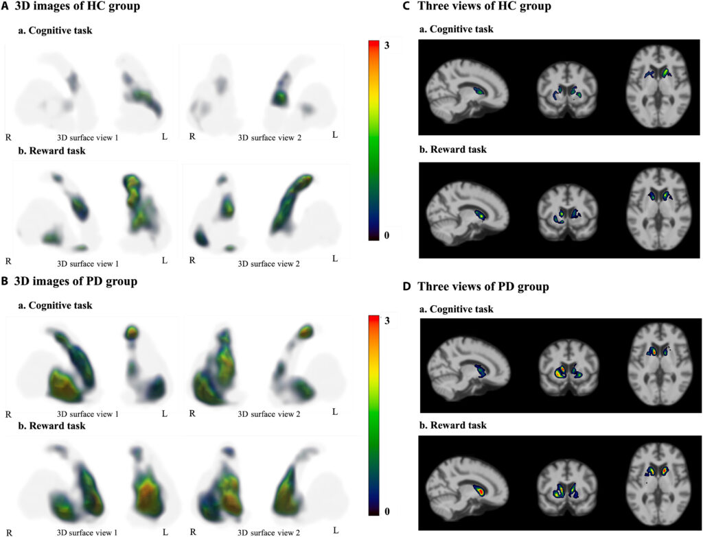
(A) 3D surface view of striatum in healthy control (HC) group. (B) 3D surface view of striatum in Parkinson’s disease (PD) group. (C) Three views of HC group. (D) Three views of PD group. The images highlight the contrast between discrete, localized dopamine release in HC and the diffuse, widespread dopamine release across extensive striatal regions in patients with PD.
Understanding compensation and its limits
While this reorganization of dopamine activity may help preserve function early in the disease, this loss of functional segregation could also contribute to Parkinson’s symptoms, including difficulty performing precise movements and multitasking.
As specialized networks become increasingly shared, the brain may lose the ability to efficiently select and coordinate individual movements or simultaneously manage motor and cognitive demands.
“Our findings show that the brain is remarkably adaptable,” says Dr. Stoessl. “But they also suggest that these compensatory changes may contribute to some of the hallmark symptoms of Parkinson’s disease as the disease progresses.”
Toward better treatments
While the results are preliminary given the study’s small sample size, Dr. Stoessl and his team note that the findings provide a better understanding of how Parkinson’s disease changes brain function and may help guide future therapies aimed at preserving the brain’s normal organization or enhancing beneficial compensation before symptoms become more severe.
“This study gives us a much richer picture of what’s happening in the brain of Parkinson’s patients,” says Dr. Stoessl. “Understanding how the brain reorganizes itself opens new opportunities to develop treatments that work with these adaptive mechanisms, rather than simply replacing lost dopamine.”
This study was made possible through funding by the Pacific Parkinson’s Research Institute (PPRI). With further funding from the PPRI, Dr. Stoessl is currently collaborating with Dr. Silke Appel-Cresswell to look at whether this functional reorganization may be modified by lifestyle interventions, including exercise.