Check out some of the papers that were recently published by DMCBH members:
Thalia Field: Cerebral Amyloid Angiopathy Imaging Markers, Radiological Boston Criteria, and Thrombolysis Outcomes in Ischemic Stroke
Journal: Stroke
Cerebral amyloid angiopathy (CAA) is thought to increase the risk of post-thrombolytic brain bleeding, but CAA neuroimaging markers and MRI criteria have not been systematically evaluated in large stroke trials. Researchers examined the association of radiological Boston CAA criteria and their constituent markers with hemorrhagic risks and functional outcomes after intravenous thrombolysis in the Alteplase compared to Tenecteplase (AcT) trial. Among 1,600 patients in the trial, 482 had suitable MRIs. Cortical superficial siderosis (CSS), the deposition of blood-breakdown products along the surface of the cerebral hemispheres, emerged as the dominant harmful neuroimaging marker. CSS burden was strongly and consistently associated with higher risk of severe hemorrhage, disability and death, making it a particularly relevant CAA marker when weighing thrombolytic risk vs benefit. Meeting radiological Boston criteria versions 1.0 or 1.5 increases hemorrhagic risk but meeting the latest 2.0 criteria does not.
Anthony Traboulsee, Roger Tam: Monitoring morphometric drift in lifelong learning segmentation of the spinal cord
Journal: Imaging neuroscience
Morphometric measures from spinal cord segmentations can serve as diagnostic and prognostic biomarkers in neurological diseases and injuries affecting the spinal cord. Automatic segmentation methods have been developed over the past few years, but it is unclear whether their predictions are stable as the model is updated with new datasets containing variable contrasts and pathologies. Researchers presented a spinal cord segmentation model trained on a multisite dataset, including 9 different MRI contrasts and several spinal cord pathologies. They also introduced a lifelong learning framework to automatically monitor the morphometric drift as the model is updated using additional datasets. Results showed that the model performs well, with a smooth automatic workflow and minimal drift between different versions of the model. The code and model are open source and accessible via Spinal Cord Toolbox v7.0.
Journal: eGastroenterology
Some people with multiple sclerosis (MS) show changes in their gut microbiota, with evidence suggesting that MS symptoms may worsen with a high-fibre diet. Researchers hypothesized that in people with multiple sclerosis whose gut microbiota are less able to efficiently ferment dietary fibres, unfermented β-fructans induce inflammation. The gut microbiota of individuals with paediatric-onset multiple sclerosis showed reduced fibre fermenting properties, and lower β-fructan consumption was observed among participants with paediatric-onset multiple sclerosis. A germ-free mouse model that was unable to ferment fibres was used to study the response to unfermented β-fructans. Results suggest that unfermented β-fructans can worsen demyelination and promote gut-brain axis immune activation. Future longitudinal studies are warranted to confirm the findings uncovered in this manuscript.
Catharine Rankin: Neuropeptides Are Involved in Elicited Reversal Speed Plasticity in C. elegans During Mechanosensory Habituation
Journal: MicroPublication biology
Caenorhabditis elegans respond to mechanosensory taps with brief reversal responses. In past research, speed of the response was averaged over the entire reversal for each tap to analyze reversal speed habituation; however, with this measure, only a modest decrement in speed was observed. Using a more detailed breakdown of reversal speed, we found that speed is most plastic early in the reversal and stable later on. Using this analysis, we found that worms with mutations in neuropeptide genes show reduced speed plasticity during the first second of reversals, indicating peptidergic signaling may be involved in reversal speed plasticity.
Helen Tremlett: Interventional studies targeting the prodromal or preclinical phase of immune-mediated disease to benefit patient outcomes: a scoping review
Journal: Journal of autoimmunity
Many autoimmune diseases such as multiple sclerosis, rheumatoid arthritis, lupus and type 1 diabetes begin silently, with early biological changes happening before noticeable symptoms appear. This review looked at studies testing whether treating people during the prodromal stage can delay or prevent full illness. Out of more than 1,400 studies screened, 23 met the criteria. The strongest evidence was for rheumatoid arthritis and type 1 diabetes. In type 1 diabetes, the drug teplizumab delayed diagnosis by more than two years on average and helped preserve insulin-producing cells. Some drugs also reduced the risk of developing rheumatoid arthritis and multiple sclerosis in high-risk individuals. Overall, these early treatments were generally safe and showed promise in slowing disease onset, but more research is needed and clearer definitions and longer follow-up will help move this prevention approach forward.
Daniela Palombo: Harmonized Protocol for Subfield Segmentation in the Hippocampal Body on High-Resolution In Vivo MRI From the Hippocampal Subfields Group (HSG)
Journal: Hippocampus
Different parts of the hippocampus, a brain region essential for memory, develop and age differently, and some are more vulnerable to diseases like Alzheimer’s. Although high-resolution MRI now allows scientists to study these smaller sections, labs have used different methods, making results hard to compare. An international team called The Hippocampal Subfields Group created a standardized, research-based guide for identifying key hippocampal subfields in brain scans. The method was carefully developed, reviewed by experts, and shown to be reliable across researchers. This shared approach will improve consistency across studies and deepen our understanding of how the hippocampus changes over time and in disease.
Martin McKeown: Low-intensity transcranial ultrasound effects on the ventral intermediate nucleus and zona incerta in Parkinson’s disease tremor
Journal: Brain Stimulation
This study tested whether a non-invasive technique called transcranial ultrasound stimulation (TUS), which uses sound waves to stimulate deep brain areas, could reduce tremor in people with Parkinson’s disease. Researchers compared two brain targets: the ventral intermediate nucleus (VIM) of the thalamus and a nearby region called the zona incerta (ZI). Stimulating the VIM improved tremor that occurs while holding a posture, but not tremor at rest. In contrast, stimulating the ZI reduced both postural and resting tremor. Brain scans showed that changes in brain activity and connectivity after ZI stimulation were linked to how much a person’s tremor improved. Overall, the findings suggest that the zona incerta may be a promising new target for treating Parkinson’s tremor.
Journal: Trials
This study tested whether a 10-week, online seated exercise program can improve mobility in people living with long-term effects of stroke who have balance problems. Researchers compared people who complete the virtual seated exercise program three times a week with a control group. They measured changes in strength and mobility (such as how many times someone can stand up from a chair in 30 seconds) as well as balance, quality of life, mood, fatigue and overall health. The study found that chair-based exercises can help improve movement and independence after stroke. Because the exercises are done while sitting, it also improves safety and therefore increase participation.
Khaled Abdelrahman: Neuroglial Function and Hormonal Modulation in Neurodegenerative Diseases: The Influence of Sex Hormones
Journal: Cellular and molecular neurobiology
Support cells in the brain, such as astrocytes, microglia, and oligodendrocytes, play vital roles in keeping brain cells healthy, regulating inflammation and maintaining connections between neurons. In diseases such as Alzheimer’s, Parkinson’s, Huntington’s and ALS, these support cells can stop working properly, contributing to ongoing inflammation, damage to brain connections, and loss of protective insulation around nerve fibers. This review highlights growing evidence that sex hormones like estrogen and testosterone influence how these brain support cells function. These hormonal effects may help explain why some brain diseases differ between men and women in terms of risk, progression and response to treatment. Understanding how hormones interact with brain support cells could lead to more personalized, sex-informed treatments for neurodegenerative diseases.
Journal: Neuron
Severe traumatic events, such as natural disasters or mass shootings, can lead to long-lasting memory problems and heightened sensitivity to reminders of the trauma, especially in women. In this study using mice, researchers explored how the hormone estrogen (specifically estradiol) affects the brain’s response to intense stress. Surprisingly, higher natural levels of estradiol in the hippocampus were linked to greater vulnerability to stress-related memory disruption. The hormone appeared to make certain genes more “open” and active, increasing sensitivity to trauma. Different estrogen receptors also played distinct roles in males and females. These findings suggest that hormone levels and biological sex influence how the brain responds to trauma, which may help explain sex differences in stress-related disorders.
Silke Appel-Cresswell: A randomized safety and feasibility crossover trial of two Mediterranean-ketogenic interventions in individuals with Parkinson’s disease
Journal: Journal of Parkinson’s disease
Researchers tested two versions of a Mediterranean-style diet designed to increase ketones (an alternative brain fuel) in people with Parkinson’s disease. One was a high-fat, low-carbohydrate Mediterranean ketogenic diet, and the other was a standard Mediterranean diet with added medium-chain triglycerides (MCT oil). Both diets were generally safe over the short term, and no serious side effects were reported. However, many participants found the diets difficult to stick to, and more than a third did not complete the full study. Surprisingly, small improvements in daily motor symptoms were seen only in the MCT-supplemented diet group—even though most people on that diet did not reach full ketosis. The results suggest these diets are safe but challenging to maintain, and future studies need better strategies to help people follow them consistently.
Mypinder Sekhon, Mark Cembrowski: Omics-based approaches for acute brain injury research
Journal: Current opinion in critical care
Acute brain injuries, such as those caused by trauma, stroke, or lack of oxygen, vary widely from person to person, and treatment options remain limited. Current care mainly focuses on supporting the brain rather than targeting the underlying damage. Researchers are now turning to “omics” approaches, which study large sets of biological information like genes (genomics), gene activity (transcriptomics), proteins (proteomics), and metabolism (metabolomics). These tools can help identify specific biological pathways involved in different types of brain injury and uncover biomarkers that signal how severe the injury is or how someone might respond to treatment. By better understanding the biology of each patient’s injury, omics research could pave the way for more personalized and effective treatments in the future.
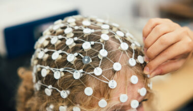
Naznin Virji-Babul: Altered resting state EEG microstate dynamics in acute concussion in adolescents
Journal: Scientific Reports
Concussions can disrupt how the brain functions, but clinicians still lack clear, objective tools to measure these changes. In this study, researchers used EEG—a test that records brain waves—to examine brief patterns of brain activity called “microstates” in adolescents within two weeks of a concussion. Compared to healthy individuals, those with a concussion showed clear differences in how long certain brain activity patterns lasted and how often they occurred. These changes suggest that concussion temporarily alters communication between large brain networks. The findings show that analyzing brain wave patterns at rest could become a useful, brain-based tool to help detect and better understand concussion.
Noah Silverberg: Prevalence and Risk of Anxiety and Depression after Concussion: A TRANSCENDENT Study
Journal: Journal of neurotrauma
Anxiety and depression are common after concussion, but they may be more widespread than previously thought. In this large study of over 1,600 teens and adults seen at concussion clinics, nearly half reported moderate-to-severe anxiety symptoms and more than 60% reported moderate-to-severe depression symptoms at their first visit. Higher risk was linked to being injured in a car crash or at work, being female, having anxiety before the injury, and experiencing sleep problems afterward. These findings highlight the importance of routinely screening for mental health concerns after concussion—especially in higher-risk patients—so that treatment and support can begin early and improve recovery.
Neil Cashman: Reliable repurposing of the antibody interactome inside the cell
Journal: Nature communications
Most proteins in the human body can be targeted by lab-made antibodies, but these antibodies usually don’t work well inside cells because they don’t dissolve properly in the cell’s fluid. In this study, researchers identified key features, especially electrical charge, that affect whether small engineered antibodies (called “intrabodies”) remain stable and functional inside cells. By redesigning parts of these antibodies and using artificial intelligence to guide the process, they were able to create highly stable, soluble intrabodies that still recognize their original targets. The team developed more than 600 intrabodies aimed at 60 different proteins, including ones linked to diseases such as cancer and neurodegenerative disorders. This work opens the door to using antibodies as tools to study and potentially treat diseases from inside the cell.
Silke Appel-Cresswell: Virtually Together: Exploring the Impact of a Low-Latency Synchronized Audio Platform on Participant Wellbeing in a Facilitated Online Singing Group
Journal: Music & Science
Group singing is known to boost mood and wellbeing, but it’s been unclear whether the same benefits occur online, especially since audio delays often prevent participants from singing together in real time. In this study, researchers compared two types of online singing sessions for older adults and people with neurological conditions: one where participants were muted and only heard the leader, and another using newer technology that allowed everyone to hear each other live. Both formats improved mood and feelings of social connection, and singing was linked to lower stress levels and less self-reported pain. However, sessions where participants could hear each other in real time led to stronger feelings of connection and enjoyment. The findings suggest that virtual group singing can meaningfully support wellbeing, especially for people who cannot attend in-person activities.
Journal: The Lancet Digital Health
After a stroke, the brain undergoes long-term structural changes that can affect movement. In this large international study, researchers examined whether certain parts of the brain appear “older” or “younger” than expected on MRI scans—a measure called brain predicted age difference (PAD)—and how this relates to motor recovery. They found that larger stroke lesions were linked to faster “aging” in the damaged side of the brain. Interestingly, the opposite side of the brain sometimes appeared “younger,” suggesting it may be adapting or compensating to help with recovery. Damage to key motor pathways and important brain networks was strongly linked to worse movement outcomes. These findings suggest that measuring the “age” of specific brain regions could help doctors better understand recovery after stroke and potentially guide more personalized rehabilitation strategies.
Vesna Sossi, Jon Stoessl, Teresa Liu-Ambrose: Exercise-Induced modulation of molecular-enriched functional connectivity in Parkinson’s disease
Journal: Journal of Parkinson’s disease
Parkinson’s disease (PD) affects multiple aspects of brain function including movement. Exercise can improve symptoms, but it is unclear how it changes the brain. In this study, researchers combined brain imaging (fMRI) with maps of key neurotransmitters to see how exercise affects brain networks in PD. The team studied 22 people with PD, including a subgroup who underwent six months of aerobic exercise, and compared them to healthy controls. Using a method called REACT, they looked at brain circuits linked to dopamine, acetylcholine, serotonin, and noradrenaline. The results suggest that exercise may help partially restore normal function in circuits disrupted by PD, especially those related to dopamine and acetylcholine. This study provides supporting evidence that exercise changes brain networks in a way that could improve symptoms and supports using molecular-informed imaging to understand these effects.
Daniela Palombo: Take me back: how the temporal relationship between cues shapes emotional memory retrieval
Journal: Psychological research
Emotional moments, like frightening or joyful events, usually happen alongside more neutral details that come before and after them. This study explored how those surrounding neutral moments become linked to the emotional experience in memory. Participants viewed short sequences of images: a neutral image, followed by either an emotional or neutral scene, and then another neutral image. Later, they were tested on how well they remembered the full sequence. The researchers found that when people were reminded of what happened after a negative emotional event, they were better able to recall specific details and the emotional intensity of the experience. This suggests that what happens after an emotional moment may play a special role in shaping how we remember meaningful life events.
Khaled Abdelrahman: Role of Endocannabinoid System Perturbation in Organophosphate-Mediated Metabolic Impairment and Neuroinflammation
Journal: Basic & clinical pharmacology & toxicology
Organophosphates (OPs), commonly used as pesticides, have been shown to adversely affect both metabolism and brain health by disrupting the endocannabinoid system (ECS), a critical regulatory network involved in inflammation, energy balance, and neural function. Chronic, low‐dose exposure to OPs can alter ECS enzymes and signalling pathways, contributing to insulin resistance, obesity and neuroinflammation. These metabolic disturbances may play a key role in the development of neurodegenerative outcomes associated with OP toxicity. This review aims to examine the interplay between OPs exposure and ECS disruption, emphasizing the ECS role in pathogenesis and its potential as a therapeutic target.
William Panenka, Alasdair Barr, Alexander Rauscher, Thalia Field: Longitudinal brain atrophy and mortality among people living in homelessness and precarious housing: A brief report of a longitudinal study
Journal: PloS one
People experiencing homelessness or unstable housing often face serious health challenges. In this long-term study in Vancouver, researchers used brain scans to track changes in brain volume over about seven years in more than 300 individuals. They found that alcohol dependence and higher cardiovascular risk were linked to faster brain shrinkage over time, and a history of moderate or severe traumatic brain injury was linked to greater brain shrinkage at the start of the study. Importantly, greater brain shrinkage was also associated with a higher risk of death during the study period. These findings suggest that addressing treatable risk factors—such as alcohol use and heart health—could help protect brain health and potentially improve survival among people living without stable housing.
Mark Cembrowski: Divergent hippocampal output via covariant local and long-range neuronal structure
Journal: Progress in neurobiology
The brain’s structure helps determine how it processes information, but scientists are still learning how small differences in cell shape affect brain function. In this study, researchers focused on a group of neurons in the subiculum, a key output region of the hippocampus involved in memory. By reconstructing individual neurons in detail and using computer modeling, they found that these cells show a coordinated pattern between their branching “input” structures (dendrites) and their long-range “output” connections (axons). This matching design appears to shape how each neuron processes and sends information. The findings suggest that even subtle differences in a neuron’s shape can influence how specific brain circuits function, revealing a new level of organization in memory-related pathways.
Kiran Soma, Annie Ciernia: Neonatal lipopolysaccharide has long-term effects on steroids and gene expression in the adult mouse brain
Journal: Hormones and behavior
Infections during early life may have lasting effects on brain development. In this mouse study, researchers examined how an immune challenge shortly after birth affected brain hormones and gene activity later in adulthood. Mice exposed to an immune trigger as newborns showed long-term changes in stress-related hormones in several brain regions, especially when they experienced another immune challenge as adults. These early infections also altered patterns of gene activity in the hippocampus, a region important for memory and stress regulation. The findings suggest that early-life infections can “program” how the brain responds to stress and inflammation later in life, potentially influencing long-term brain health and behavior.
Mahmoud Pouladi: TSC2 GAP Domain V1646Cfs*7 Variant Alters Protein Stability and Interaction Networks in Tuberous Sclerosis Complex
Journal: Neurology Genetics
Tuberous sclerosis complex (TSC) is a genetic condition that can cause epilepsy and developmental delays. In this study, researchers examined a child with severe, treatment-resistant seizures who had a newly identified mutation in the TSC2 gene. They found that this mutation produces a shortened version of the TSC2 protein that still reaches the right place in the cell and interacts with key growth pathways, but breaks down more quickly than normal. Further analysis showed that the altered protein affects important processes such as RNA regulation and how cells manage their energy-producing mitochondria—pathways that are also linked to autism and epilepsy. These findings help explain how this specific mutation may lead to disease and point toward more personalized treatment approaches for TSC and related genetic disorders.
Mypinder Sekhon: Hyperoxaemia in acute brain injured patients: too much of a good thing?
Journal: Intensive Care Medicine
The brain is highly sensitive to oxygen levels: too little oxygen (hypoxaemia) can quickly cause irreversible damage, but too much oxygen (hyperoxaemia) may also be harmful in with acute brain injuries, including cardiac arrest, stroke, traumatic brain injury, and hemorrhages. High oxygen levels or large fluctuations can worsen outcomes by causing oxidative stress or constricting blood vessels, reducing blood flow to already vulnerable brain tissue. While giving extra oxygen is essential to prevent hypoxaemia, studies show mixed results on whether conservative oxygen strategies improve survival and neurological recovery in the ICU. Large trials currently underway aim to clarify the safest and most effective oxygen targets, highlighting that balancing oxygen to avoid both deficiency and excess remains a critical, unresolved question in neurocritical care.
Noah Silverberg: Network models of symptoms following mild traumatic brain injury: A systematic review and meta-analysis
Journal: Journal of the International Neuropsychological Society
After a mild traumatic brain injury (mTBI), people often experience a mix of cognitive, emotional, and physical symptoms, but how these symptoms interact isn’t fully understood. This study combined data from six large samples (5,776 participants) to create the first pooled “symptom network” for post-concussion effects. The analysis showed that cognitive problems—especially trouble concentrating and slowed thinking—were the most central symptoms, linking closely to emotional and physical complaints. However, the study also found large differences between individual studies, suggesting that post-concussion symptoms can vary widely between people. These results highlight the importance of focusing on cognitive symptoms and suggest that personalized, patient-specific approaches may be needed for effective rehabilitation after mTBI.