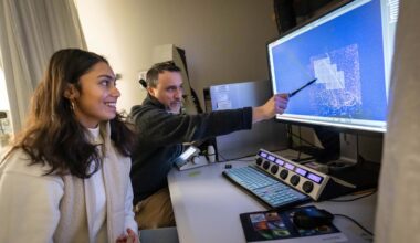
The Dynamic Brain Circuits research excellence cluster’s new iMAP (in vivo Mesoscale Assessment of the neuroProjectome) project is using advanced imaging tools to assess and manipulate brain circuits in animal models of human brain disorders.
Led by Dr. Tim Murphy and managed by Research Associate Jeff LeDue, iMAP discoveries will lead to new treatments, called circuit-based therapeutics, which can be personalized to an individual’s needs. These discoveries will be enabled by iMAP’s high-resolution imaging of the function and structure of projections between widely-separated areas of the brain, bridging the current gap between preclinical studies and patient imaging.
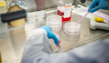
With support from the Canadian Foundation for Innovation and BC Knowledge Development Fund, the iMap project has provided Djavad Mowafaghian Centre for Brain Health researchers with over $3 million in new equipment and resources in 2022 to date, including a Lattice Light Sheet Microscope (one of two in Canada), two fiber photometry systems and a multiphoton mesoscope (one of five in the world).
A new review paper published in Neuron today (written by Murphy, LeDue and Wang) highlights the importance of using multiscale imaging techniques, including those within the iMAP portfolio, to study the structure and function of neurons in different parts of the brain of mouse models. These animal studies are essential for understanding various neurodegenerative disorders and developing new therapeutics.
Some of the emerging imaging techniques discussed in the review include:
- Functional Ultrasound (fUS): fUS enables functional MRI-like imaging, but with more portability and lower cost. The fUS system allows for structural imaging of the brain and quantification of cerebral blood volume and flow.
- Multiphoton / Ultra Wide-field Multiphoton: Multiphoton, while more invasive, provides a large field of view and allows for a more direct examination into behavioural activity and cellular imaging of functional signals from specific neurons over multiple brain regions.
- Wide field epifluorescence: Wide field cortical surface epifluorescence provides a simple, reduced resolution large field-of view method and is useful for mesoscale circuit assessment.
- Miniscopes: Miniscopes allow for tissue penetrating wide field imaging but are more invasive and require surgical implantation.
- Fiber Photometry: Fiber photometry allows for less invasive imaging and enables optical recording from genetically targeted sub-populations of neurons throughout the brain during freely moving active behaviour.
- Lattice Light Sheet Microscopy: With its ability to image both dendrites and axons of individual neurons, electron microscopy is the gold standard in the study of structural connectivity within fixed tissues.
Each of the imaging techniques highlighted above can be useful in various animal models for different diseases. Imaging blood flow is especially useful in stroke models and can show microvascular abnormalities and cellular changes post-stroke. In Alzheimer disease models, these techniques can be used to examine blood flow, neuronal activity, circuit connectivity and the structure of amyloid plaques. For Huntington disease models, neuronal activity can be measured during behavioural tasks, such balance and motor tasks, while in Parkinson disease models, imaging can help to track neurotoxins and changes in genetic variants.
With these multiscale techniques, precise cell and circuit manipulation is possible, which can ultimately lead to new therapeutics that can restore motor and cognitive function, as well as psychological wellbeing to patients with neurological diseases.
However, all of these imaging tools also result in a large amount of data. The use of open source repositories and analysis tools, along with standardized procedures and documentation, is important to the future of open neuroscience.
By employing the principles of open science, the iMAP project will accelerate discovery and facilitate collaborations between scientists and clinicians at the Centre and beyond. Together, these new resources will allow researchers who are working to understand how circuitry in the brain is impacted by diseases, injuries and illnesses, to examine brain function and structure at sizes and times scales that were not previously accessible.