When we think about the immune system, we often think about its role in fighting infections and protecting us from disease. But some immune cells have another important job: helping build and shape tissues throughout the body.

Dr. Rosin
Recent research from Dr. Jessica Rosin’s lab is uncovering how colony-stimulating factor 1 receptor (CSF1R)-expressing immune cells help guide the formation of bones, teeth and facial structures during embryonic development.
“CSF1R is well known for its role in immune cell survival, allowing us to target this signalling pathway to study the important role immune cells play in the environment that growing tissues rely on,” says Dr. Rosin, a Tier 2 Canada Research Chair in Immune Regulation of Developmental Programs, Assistant Professor in UBC’s Faculty of Dentistry and member of the Djavad Mowafaghian Centre for Brain Health.
CSF1R is found on several types of cells, including macrophages and osteoclasts. Macrophages help clear damaged cells and debris, while osteoclasts break down and remodel bone. Dr. Rosin’s research suggests that these cells have important roles during development that extend beyond their traditional functions.
A receptor with a role in bone development
In a recent study published in the Journal of Developmental Biology, Dr. Rosin and her team examined the role of CSF1R in developing limbs. To investigate what happens when CSF1R signalling is disrupted, pregnant mice were given PLX5622, a drug that inhibits CSF1R. The result was significantly reduced CSF1R-expressing cells in developing limbs and the elimination of detectable osteoclast activity in the embryonic bones.
In early stages of development, the limbs appeared largely normal. However, by birth, all of the limb bones examined were shorter than those of untreated mice, and some bones in the heel were missing or underdeveloped. These findings suggest that CSF1R-dependent cells are important during a specific window of development, even when changes are not immediately visible.
“Development can look normal at one stage, while changes happening behind the scenes have consequences that only become apparent later,” says Dr. Rosin.
The research team also found differences between mouse strains, suggesting that genetics can influence how strongly the developing skeleton responds to changes in CSF1R signalling.
CSF1R helps in shaping the skull and face
Dr. Rosin’s research also shows that CSF1R plays an important role in forming the skull and other structures of the face. In a study published in Development, the Rosin lab used PLX5622 to block the receptor during pregnancy in mice, which resulted in 50 per cent depletion of macrophages and a complete loss of osteoclasts.
This caused a range of changes to the skull and face, including a more rounded or domed skull and abnormalities in the jaw, palate, ear bones and other parts of the craniofacial skeleton. Changes were also found in the cranial sutures, which are the joints between the bones of the skull that allow the skull to grow.
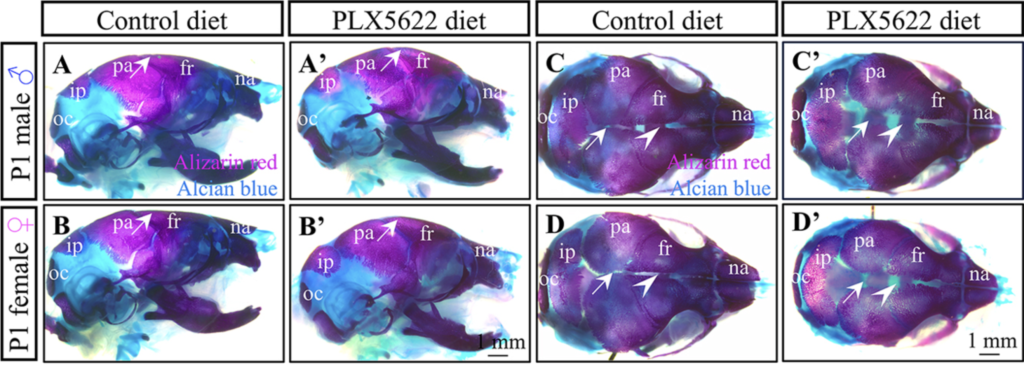
Prenatal exposure to PLX5622 disrupts formation of the skull. Arrows in A-B′ indicate skull doming and arrows in C-D′ show suture impairments.
More importantly, the effects were not limited to bones. Disrupting CSF1R altered the signalling molecules involved in communication between cells and reduced the growth of neural crest cells, a population of cells that contributes to many structures in the face and skull. These findings suggest that CSF1R-dependent cells can influence development indirectly by helping shape the signals and cellular environment around them.
“Macrophages and osteoclasts are part of a much larger developmental environment,” says Dr. Rosin. “Changing these cells can affect the signals that other cells receive, with lasting effects on how tissues form.”
The environment around developing teeth
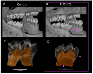
CSF1R inhibition causes abnormal shape changes to first molars, including a distinct bulge of enamel (as shown in B).
A similar pattern emerged in research on teeth development. In a separate study published in the Journal of Developmental Biology, Dr. Rosin and her team found that when CSF1R was blocked during embryonic development, mice developed unusual changes in their incisors and first molars, including abnormal shapes and folds. However, CSF1R was not detected in the main cells responsible for forming the teeth. Instead, CSF1R-dependent cells were found in the surrounding tissues.
This suggests that the receptor may indirectly influence tooth formation. Osteoclasts, for example, help remodel the bone surrounding a developing tooth, potentially creating the space and environment needed for the tooth to take shape. The timing of CSF1R activity also appears to be important as blocking CSF1R after birth did not produce the same abnormalities in continuously growing adult incisors.
“We found that the environment surrounding a developing tooth can have a major influence on its final shape,” says Dr. Rosin.
Connecting bones, muscles and the immune system
More recent work from the Rosin lab, published in Cells & Development, has also shown how CSF1R affects skull, jaw and facial muscles in newborn mice.
Using high-resolution imaging, the team found that prenatal CSF1R inhibition altered the shape of the skull and jaw, as well as muscles involved in chewing and tongue movement. Some of the muscle changes closely followed changes in the bones where those muscles attach. The findings suggest that changes to the skeleton may influence the shape and growth of nearby muscles, although Dr. Rosin notes that CSF1R-dependent cells within the muscles could also contribute directly.
Importantly, the changes were already apparent at birth and some became even more pronounced after birth, even though CSF1R signalling resumes once the drug is removed. This suggests that changes occurring before birth can establish a developmental trajectory that continues after the initial disruption.
“What’s particularly interesting is how closely connected the changes in bone and muscle are,” says Dr. Rosin. “It suggests that early changes in the tissue environment can influence how these structures grow after birth.”
A bigger picture of development
Together, these studies point to a broader role for CSF1R in shaping the environment in which tissues form. The receptor helps support macrophages and osteoclasts, but the effects of these cells extend beyond traditional immune and bone-remodelling functions. During embryonic development, they appear to help create the conditions that allow bones, teeth and muscles to form properly.
“Our findings highlight how closely connected tissues are during growth,” says Dr. Rosin. “Changes in the cells surrounding a structure can ultimately shape its formation, even when those cells are not directly involved in building it.”
Understanding how immune cells contribute to development could provide new insight into how disruptions before birth may lead to neurological and skeletal abnormalities, while revealing the broader roles CSF1R plays throughout the body at different stages of life.