Magnetic resonance imaging (MRI) scanners can be programmed to measure myelin, which is the insulating layer that protects neurons and is a key indicator of numerous neurological disorders. However, myelin imaging is extremely slow, traditionally requiring the MRI scanner to collect huge amounts of data. UBC researchers have developed a new technique that collects a tiny fraction of the data while providing higher-quality myelin measurements.
This new method developed by Dr. Adam Dvorak, under the supervision of Faculty of Medicine Associate Professor Dr. Shannon Kolind, is called the Constrained, Adaptive, Low-dimensional, Intrinsically Precise Reconstruction (CALIPR) framework. A recent study published in Science Advances examined the use of the CALIPR framework specifically in the context of myelin water imaging (MWI), which is a non-invasive technique used in quantitative MRI to measure the amount of myelin in the human body.
“Using this CALIPR technique, we can collect a tiny fraction of the imaging data — less than five per cent — but still reconstruct high-quality images,” says Dr. Dvorak, the study’s first author and recent PhD graduate in the Kolind lab. “Less data means less time lying in the scanner — in this case, more than 20 times less!”
The CALIPR technique allows for the acquisition of advanced, high-resolution quantitative maps of the human brain in 7.5 minutes, instead of 3 hours, making myelin imaging approximately 25 times faster than a conventional full MRI scan.
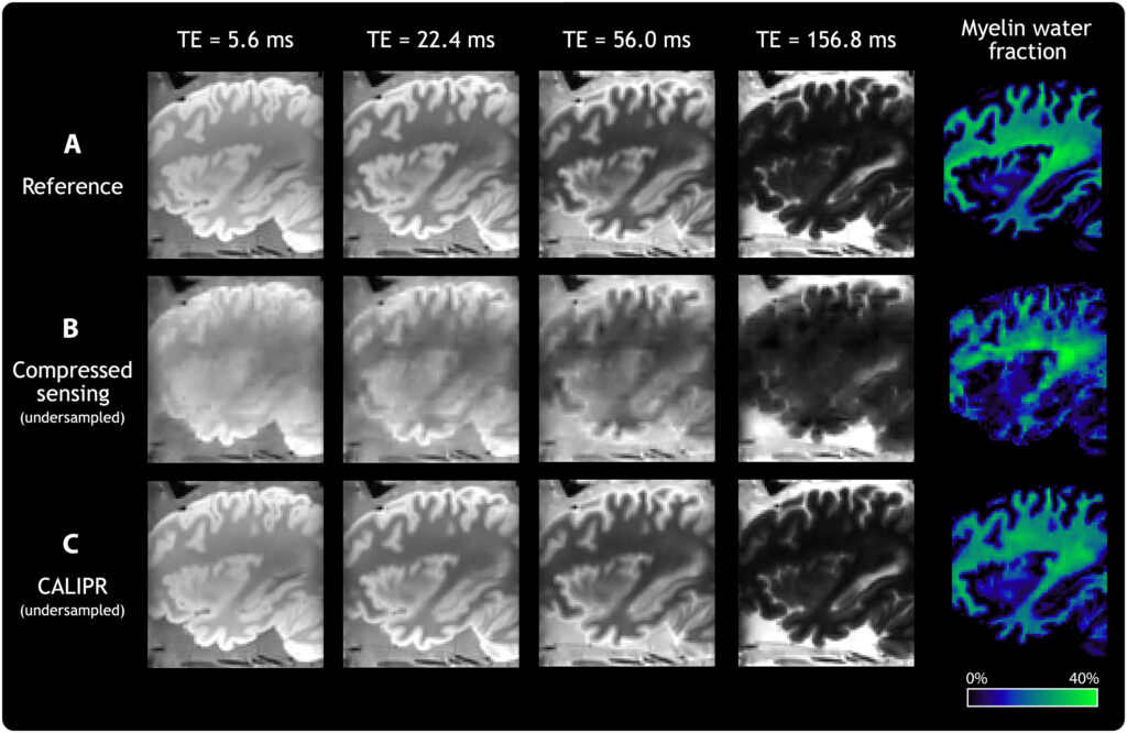
This figure compares a “gold standard” reference acquisition (row A) to the current state-of-the-art technique for accelerating MRI acquisition (row B) and the new CALIPR technique (row C). While the “compressed sensing” and “CALIPR” images were both generated using a tiny fraction of the imaging data compared to the reference, making the imaging process much faster, it is clear the CALIPR images are much more detailed.
The new framework has demonstrated excellent precision in quantifying myelin content, with low variability in measurements for both the brain and the spinal cord. Compared to existing techniques, CALIPR is much more efficient and can potentially improve the detection of demyelinating diseases such as multiple sclerosis.
“If we compare an MRI image reconstruction to solving a multiple choice question, this technique does the equivalent of eliminating most of the incorrect options, making it much easier to solve.” Dr. Dvorak explains.
This technique, available for three different MRI manufacturers including a small, portable MRI scanner, may have broader applications in other quantitative MRI techniques and help revolutionize the treatment and care of a wide range of neurological disorders.
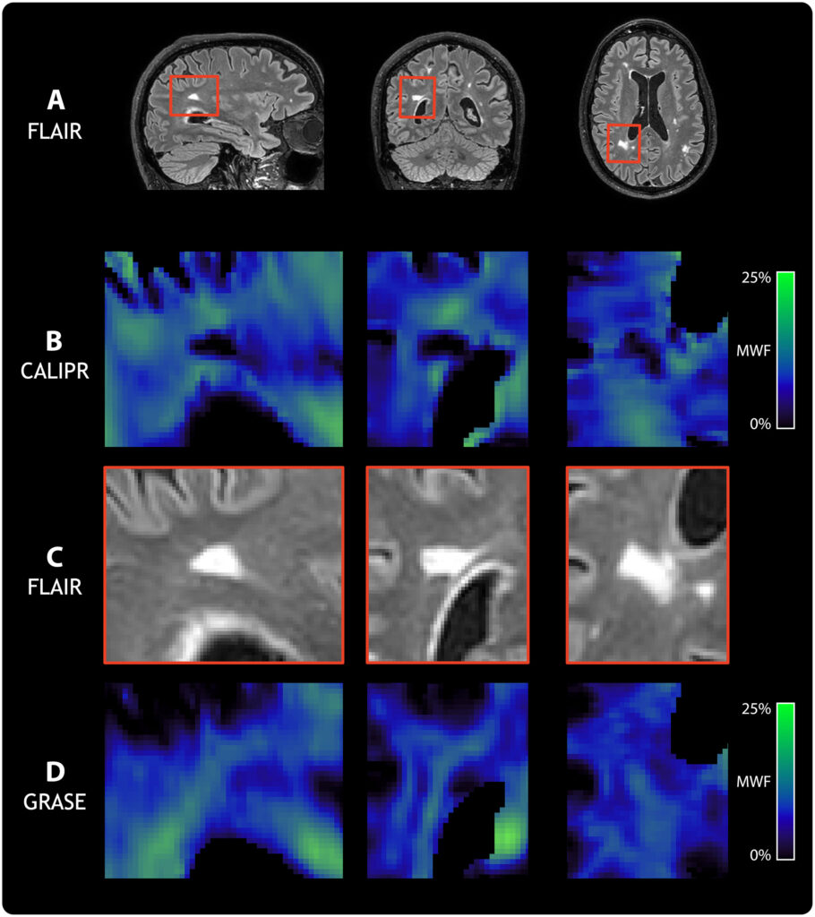
This figure depicts a high resolution MRI image showing anatomy (row A) with a closeup of tissue affected by disease (row C) to compare to advanced quantitative MRI maps of tissue structure acquired with the CALIPR technique (row B) and the current state-of-the-art GRASE technique (row D). It is clear the CALIPR technique (row B) provides higher quality, higher resolution maps that better identify (and quantify) damage to the tissue at the location of the bright lesion (visible in row C).