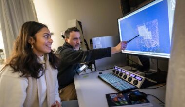
Pictured: Elisa York at the Djavad Mowafaghian Centre for Brain Health. Image credit: Paul Joseph/UBC.
Necessity is the mother of invention, and Elisa York, a PhD student in Dr. Brian MacVicar’s lab found that in addressing a need in her own research she could solve a larger problem for other researchers studying microglia (the brain’s immune cells).
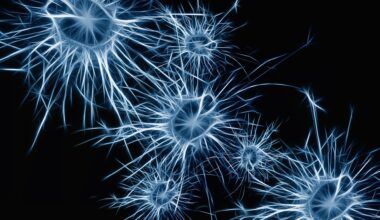
Microglia are active, shape-shifting cells that change their appearance based on their level of activity. They are small cells, each with probing processes (arm-like extensions) that monitor brain tissue for threats in the form of infection or injury. When active, microglia processes extend outward and work quickly to mitigate cellular and tissue damage; when compromised or deprived of oxygen, microglia retract their processes.
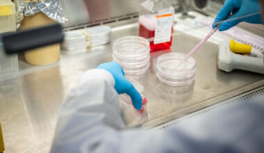
Because the appearance of individual microglia is variable depending on function, it can be hard to capture detailed morphology (observable structural features of the cell). Researchers use microglia morphology to understand their function within their environment, so it’s important that labs be able to produce reliable, reproducible images from their imaging data.
A new tool from York and colleagues offers an opportunity to standardize microglia morphology analysis, automating the process and eliminating the variability that comes from each lab having a different approach to manually interpreting two-dimensional images comprised of composite imaging data.
So excited to finally share 3DMorph! It’s a free download on GitHub – check it out, and enjoy your time while it analyzes cells for you! Contact me if you have questions. Thanks to @JeffLeDue, LP Bernier, @brian_macvicar, and #eNeuro for making it possible! https://t.co/oIdi1iIF4v
— Eli York (@elimyork) DECEMBER 12, 2018
The tool, called 3DMorph, offers a scalable, interactive, automatic script that models cell data three-dimensionally. 3DMorph is adaptable to a range of imaging needs, and doesn’t rely on staining; York hopes that it’s an approachable resource for users in a variety of fields.
“The benefit to 3DMorph is that it offers flexibility for individual labs to customize, but it also reduces time spent manually tracing cell features and the margin of error associated with that approach,” says York. “It also produces reproducible images; if one lab shares their images and parameters with another lab, they’ll get the same results – we wanted to ensure the tool is not subjective or vulnerable to user biases.”
3DMorph is an open-source tool available for free on GitHub. A detailed overview of the tool was published this week in the journal eNeuro.
For York, there was a gap between the techniques available to her for her research and what she needed in order to conduct her research more effectively. York is currently working to understand the gut-brain connection, especially how changes in diet affect the gut microbiome, and how that in turn affects microglia function and activity. She will complete her PhD in the Graduate Program in Neuroscience in 2019.
Access 3DMorph at github.com/ElisaYork/3DMorph.